Gastroscopy
A gastroscopy or ‘upper GI endoscopy’ allows the endoscopist to examine the top part of the digestive system, that is, the oesophagus (gullet), stomach and duodenum (the first part of the small intestine).


Flexible sigmoidoscopy and colonoscopy
These procedures are used to examine the rectal and colonic mucosa to determine the most appropriate medical therapy.
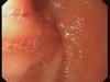
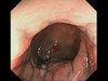
Patients require flexible sigmoidoscopy if only the distal (30-40 cm) of colon needs review. Colonoscopy is preferred if the more proximal colon requires assessment, for example if this has never been done previously; or if proximal colonic disease has not been reviewed recently (usually > 1y), especially if the appearances could result in a change in therapy e.g. anti-TNF prescription. Two biopsies should be taken from five sites including the distal ileum and rectum, and separately biopsies taken and transported in normal saline for tuberculosis (TB) culture in acute, severe colitis. Crohn’s disease is characterised by cobble stoning and discontinuous inflammation, and perianal lesions. Severity markers include deep ulceration, or submucosal ulceration affecting > ⅓ of a colonic segment (right, transverse or left colon). The scoring of endoscopic activity is mainly limited to clinical studies, although the simple endoscopic score is both straightforward to use and validated.
During colonoscopy, the risk of a perforation is about 1 in 800 and bleeding about 1 in 1500. If a polypectomy is performed, the risk of a perforation is about 1 in 600 but the risk of bleeding about 1 in 50 to 1 in 100, although this is usually mild. Sedation causes mild breathing problems in about 1 in 200.
During flexible sigmoidoscopy, there is a very small risk (1:15,000) of haemorrhage or perforation. Sedation causes mild breathing problems in about 1 in 200 cases sedated, although most patients are not sedated.


Endoscopic balloon dilation
Technique
Full bowel preparation is required. The procedure should be performed with radiological screening unless dilating short uncomplicated strictures that can be completely visualised. Stricture ≤4 cm are associated with better surgery-free outcomes.
Outcomes
Up to 90% of dilations are successful, although 1-3 dilations may be required. About 60% of patients remain symptom free at 3 years.
Risks
Perforation requiring surgery occurs in up to 5% cases, so appropriate case selection for dilation vs. stricturoplasty important.

Double and single balloon enteroscopy
Used as 2nd line procedure to diagnose Crohn’s disease (usually histologically); or provide endoscopic therapy, especially in those deemed high risk for surgery or at risk of the short-bowel syndrome.
Technique
Overnight fast required (with full bowel preparation for retrograde procedure). General anaesthesia often used, especially with anterograde procedures. The procedure takes 1-2 h. Usually visualizes 200-300 cm distal to the ligament of Treitz; and maximally 100-200 cm proximal to the ileo-caecal valve, although adhesions often limit retrograde views to 15cm of ileum.
Outcomes
The procedure is usually successful, with a diagnosis made in about 60% of patients. The retrograde approach is more difficult, and ileal intubation fails in up to 30% of cases.
Risk
Serious complications (e.g. pancreatitis, perforation, bleeding, aspiration pneumonia) occur in about 1% (up to 5% in those undergoing procedures).


Capsule endoscopy
Most experts utilise capsule endoscopy to diagnose Crohn’s disease only when clinical suspicion remains despite normal ileo-colonoscopy and small bowel cross-sectional imaging. It is occasionally used, in selected patients, to assess disease activity.
Technique
Use a patency capsule if normal cross-sectional imaging unavailable.
Outcomes
Normal capsule endoscopy has a very high negative predictive value for excluding Crohn’s disease. The overall detection rate in patients with known or suspected Crohn’s disease is ≈55% (more sensitive than other diagnostic modalities for small bowel disease), although the procedure is not specific as mucosal breaks and erosions occur in >10% normal subjects.
Risk
All patients with known or suspected Crohn’s disease should have prior small bowel imaging or (preferably) a patency study, as the risk for retention is high (≈10% and ≈2% respectively). Retained capsules may require surgery or balloon enteroscopy to be removed.